
Low-Dose X-Rays Energize New Compounds to Eliminate Brain Cancer Cells
Mice with brain tumors demonstrated double the survival rate following treatment with these new compounds
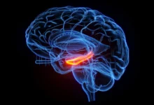
Researchers from Nanyang Technological University, Singapore (NTU Singapore), led by Prof. Pu Kanyi, have developed a groundbreaking method to treat glioblastoma, the most common type of brain cancer among adults. This new technique utilizes a substantially lower dose of X-rays compared to traditional radiation treatments, aiming to minimize side effects while effectively targeting cancer cells.
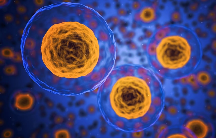
Globally, over 300,000 people are diagnosed with glioblastoma annually. This aggressive brain cancer originates from brain cells and, if untreated, spreads rapidly. The average survival time post-diagnosis is approximately 18 months. Traditional treatment options, such as radiotherapy, often lead to collateral damage to healthy cells, causing side effects like nausea, hair loss, and memory problems.
Radiodynamic therapy has emerged as a promising alternative, where patients receive specially designed compounds that generate cancer-killing free radicals when activated by X-rays. This method requires significantly lower X-ray doses—only about 20-30% of conventional radiotherapy doses. However, existing radiodynamic therapy compounds, which contain heavy metals, lack precision in targeting cancer cells and may harm healthy cells.
Prof. Pu’s team has developed a novel compound called the Molecular Radio Afterglow Dynamic Probe (MRAP), composed of biochemicals and iodine, free from heavy metals. This innovation aims to address the limitations of current radiodynamic therapy by ensuring precision in targeting cancer cells while reducing side effects.
In experiments with mice, MRAPs were directly injected into brain tumors and activated with X-rays. Remarkably, the required X-ray dosage was over six times lower than current methods. The MRAPs absorbed the X-ray radiation, generating cancer-killing free radicals only upon encountering a specific enzyme abundantly produced by brain tumor cells. This precision prevented the activation of MRAPs in normal cells, eliminating unintended side effects.


The NTU team’s experiments showed that MRAPs effectively halted tumor growth in mice, doubling their survival time from 37 to 76 days compared to untreated mice. Importantly, treated mice exhibited no tissue damage or weight loss, and the MRAPs were eventually excreted through urine and feces.
Prof. Pu highlighted the potential benefits of this method, stating, “We used very low dosages of X-rays and cancer-killing MRAPs. Also, the anti-cancer compounds were active only in the brain tumor and not healthy cells. So, we expect our treatment method to be safer and have fewer side effects than existing ones.”
A patent has been filed for MRAPs, and Prof. Pu’s team is in discussions with potential investors. The findings from this study have garnered attention from experts like Professor Marc Vendrell from The University of Edinburgh, who praised the research as a significant advancement in combining X-rays and light for precise tumor treatment.
Looking ahead, Prof. Pu’s team plans to enhance MRAPs to further improve their targeting capabilities and incorporate cancer-curbing functions such as immunotherapeutic abilities. These enhancements aim to activate the immune system to combat cancer cells and prevent recurrence.
Broader Implications
Earlier research by the team, published in Nature Biomedical Engineering in December 2022, demonstrated that another type of anti-cancer compound developed by the scientists, energized by ultrasound instead of X-rays, successfully inhibited new cancer cell growth in mice. This finding was linked to the activation of the mice’s cancer-fighting immune cells.
The development of MRAPs marks a significant milestone in brain cancer treatment, offering a safer, more precise alternative to traditional radiotherapy. With ongoing research and potential clinical applications on the horizon, this innovation holds promise for improving the quality of life and survival rates for glioblastoma patients worldwide.
Details of the study can be found in “Molecular radio afterglow probes for cancer radiodynamic theranostics” in Nature Materials (2023) and “Nanoparticles with ultrasound-induced afterglow luminescence for tumour-specific theranostics” in Nature Biomedical Engineering (2022).
References:
- Nature Materials. “Molecular radio afterglow probes for cancer radiodynamic theranostics.” Nature Materials News Release, 2023. Available from: https://www.nature.com/articles/s41563-023-01659-1.
- Nature Biomedical Engineering. “Nanoparticles with ultrasound-induced afterglow luminescence for tumour-specific theranostics.” Nature Biomedical Engineering News Release, 2022. Available from: https://www.nature.com/articles/s41551-022-00978-z.